Wednesday, October 19, 2011

Organization of the body

The anatomical position is the basic position of to decribe. The characteristics are body erect, feet slightly apart,palms facing forward, and the thumbs pointed away form the body.
This picture shows the anatomical position.

The first directional term I am going to discuss is Superior and Inferior. Superior is up toward the head or the upper part of the body. For example the chest is superior to the knees.
Inferior in the opposite of superior so it is away from the head and towards the bottom part of the body. For example the toes are inferior to the belly button. Anterior is toward the front of the body or in front. An example would be the belly button is anterior of the spine. The opposite of Anterior would be Posterior. Posterior is towards the back of the body or in the behind. The heart is posterior to the breast bone would be an example of this. Medial is going towards the middle of the body so the heart would be medial to the shoulder.

Lateral is away from the medial or middle of the body so it would be going out towards the arms or shoulders. The fingers would be lateral to the heart is an example. Proximal is closer to the origin of a body part or an limb. Like the shoulder is proximal to the elbow. Distal is going farther away from the the origin like for example, the shin is distal to the knee. Superficial is toward the the body surface. An example is the skin is superficial to the skeleton. Deep is away form the body surface and is more toward the inner organs of the body. The Kidneys are deep to the skin would be a prime example of this.
The axial view of the body is the head neck and trunk area. This is the top of the body as seen in the picture above.

There are also Appendicular appendages or limb. One of these limbs would include the Brachial which is the arm.

The next classification of the body would be the body planes or the ways the body can be split. The first one would be sagittal which divides the body into left and right parts. If you were to cut a person in half using this plane you would have the left ear, eye,leg , and arm on one side and the right side of everything on the other side.

Frontal or Coronal Plan divides the body into anterior and posterior parts. This would be dividing the body from front to back. This will be shown wonderfully from the picture seen below.


Transverse or horizontal plane divides the body into superior or inferior parts. Simply put it is dividing the body from top and bottom. The last plane is the Oblique section and this is simply cuts that are made diagonally.

The next part of the body is the body cavities. The Dorsal cavity protects the sensitive nervous system and is divided into more subdivisions. The first division is the cranial cavity that is inside the skull and is what holds the brain. The other subdivision is the vertbral cavity and that is what holds the spinal cord.

This photo shows both of the Dorsal cavity subdivisions, the cranial and vertebral. As you can see the cranial cavity is on the head and is where the brain is held. the vertebral is on the back and is what holds the spine.
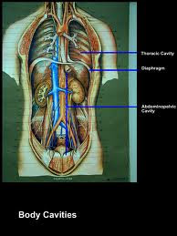
The ventral cavity holds the internal organs and just like the Dorsal cavity, has two subdivisions the thoracic and abdominopelvic.

From this picture you can infer that the ventral does indeed hold the internal organs and you can also see the two subdivisions.

Connective tissue

There is another type of tissue and it is the Connective Tissues. Just like epithelial tissues, Connective tissues are also found all throughout the body. These tissues have a variety of different duties around the body. The main job of these tissues is to provide support and protection, and is the framework for the body. For example when you are first setting up for a house you put up the framework for it made out of wood. The wood framework of the house is a good example because it is sturdy, protects the house and of course provides support. The connective tissues also have other jobs like storing fat producing blood cells and helps repair tissue damage.
One way connective tissues are different is that cells inside the tissues are more spread out than the epithelial cells. Also in connective tissues you will find extracellular matrix between the cells. This matrix is made up of a ground substance and protein fibers. The matrix can either be a solid or a fluid, or in between.
There are different types of cells included in connective tissues and the first one is fixed cells. These cells stay in a specific area of connective tissue for a certain length of time. There are also cells like macrophages that are wandering cells the move throughout the tissue usually due to and injury. Fibroblasts are another type of cells and they help produce fibers by secreting proteins into the matrix.
Macrophages are very common throughout the connective tissues just like the fibroblasts.. The macrophages are usually attached to the fibers but can move about on their own. These macrophages are important to carry on phagocytosis often police the tissues and get rid of unwanted particles. This of course is important in the bodies protection against sickness and infection.


Mast Cells are largely used throughout the connective tissues. They are almost always near blood vessels and because of this they release heparin that helps prevent blood clots. This is very crucial and without it the body would not be able to live.

Now I am going to go into the Connective tissue fibers. The first one I am going to talk about is Collagenous fibers. These are thick threads of collagen which is a protein. They are grouped in long parallel bundles and are very strong. The two big Connective tissues however, is the Ligaments and Tendons. A ligament is a fiber that connects bones to the other bones and tendons are what keep the muscles onto the bones.


Areolar Tissue are mainly fibroblasts which are seperrated by a gel like substance that has many elastic fibers. This tissue keeps the skin on the organs where there are many blood vessles.

Adipose Tissues are easy to remember to because they are pretty much just fat. These are formed when a lot of other cells store droplets of fat in their cytoplasm. As the cells get bigger they no longer look like fibers but instead they become a fat when their nuclei is pushed to the side. These joints are pillows for certain joints and organs so they are protected. Also right beneath the skin the fat cells store energy. The amount of these cells varies by the diet of the person. For example a marathon runner probably would not have as many adipose cells as would an overweight couch potato.

Homeostasis Lab

Tuesday, October 18, 2011

Epithelial Tissue

Epithelial Tissues are found all over the body. They mostly cover body surfaces, organs, and the inner lining of body cavities. These cells divide very often, causing injuries to heal quickly.  Epithelial cells are all tightly packed so they are protected very well. Epithelial Tissues are divided into many different classifications including, squamous, cubiodal and columnar. They are also divided between simple and stratified. These tissues, however, do not have blood vessels. On the other hand, connective tissues which are under these epithelial tissues do have blood vessels.

One type of Epithelial is squamous. These cells are flattened and make a smooth surface usually used for forming the walls of capillaries and membranes. To me they remind me of a smooth tile surface that lines the walls of a house or the floor. However, since the squamous cells are so thin they can be damaged or destroyed fairly easily. From this picture you can see the shape of the squamous cells.

You can see that they are thin and smooth, like the tile surface I explained earlier. It would make sense from seeing this picture that these cells would form as a wall inside the body.

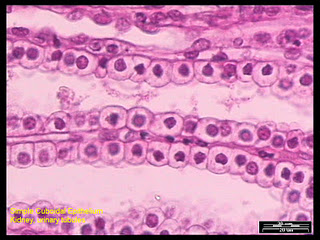
The next type of cell is the Cuboidal cells. Just saying the name of the cell can help you determine what this cell looks like. These cells are simple cube shaped cells that help with secretion and absorption of a material. In the middle is usually a large spherical nucleus.  You can usually find these cells in the kidney tubules and in the small portions of small glands.
This photo shows the shape of these unique cells. From the picture you can see that the cells are indeed cube like and have a large circle nucleus.


Columnar is the next type of cell and its name also helps reveal the shape of the cell. Columnar cells are tall cells that almost look like large columns; also the nuclei in the columnar cells are oval shaped. Some of these cells have cilia which are constantly moving. The cilia help in situations like for females moving the egg cell through the uterine tube to the uterus.


This picture shows that the long columnar cells. These cells look like large tall columns and on top of the cells, are the hair looking cilia.  The main purpose of the columnar is to absorb nutrients from digested food.
The next Epithelium is the Transitional Epithelium.   Transitional epithelium is used to change in response to increased tension. It is found in the inner ling of the urinary bladder and in the ureters. When the wall of one the organs reduces in size or contracts the tissue is usually made up of layers of cuboidal cells. Pseudo stratified Columnar is a single layer of cells that are all at different heights. Many of these cells do not even reach the free surface.  The nuclei are never in the same place they are always seen in different layers. These cells main purpose is for the secretion of mucus.


This is an example of Pseudo stratified columnar. As you can see the nuclei are in different layers.
All of these cells are split into another two division being simple and stratified.  Simple is defined usually by only a single layer of cells while as stratified cells usually have several layers of cells. Stratified cells are usually not as common as simple Epithelia cells, but they are equally as important.


The photo above shows a stratified squamous epithelia cells. As you can see there are a several layers of cells all piled up onto each other.